Technology
Digital Mammography
Optimized AEC
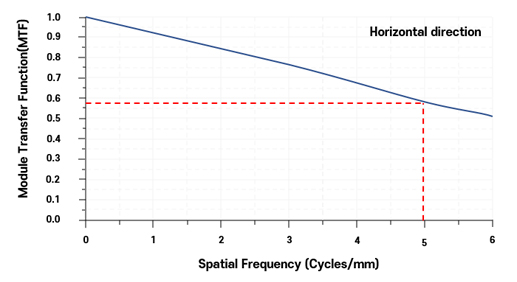
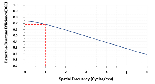
AEC is the optimal X-ray control software which is developed to minimize dose of the amount of exposure. It is an optimized algorithm that calculates the quality and dose of the amount of exposure to ensure the contrast and brightness required for the reading. Mammography requires the soft tissue of a woman to be taken differently from ordinary x-rays, which are used to photograph bones. Therefore, it is possible to reduce the dose by carefully selecting the quality of the radiation (kVp) and minimizing the dose (mAs). Low doze acquisition is implemented to guarantee the performance of MTF and DQE.
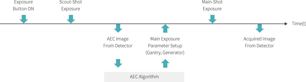
AEC images were acquired using a fast image acquisition algorithm developed for the purpose of acquiring images quickly. For this purpose, each block of the AEC image was composed of 128x128 pixels. In addition, X-ray irradiation conditions optimized for each situation were subdivided by breast thickness, shooting mode, and focal spot, and a perfect exception condition was handled for patient protection. Implanted breast is an example of this, in which case the security off process is performed and the detection algorithm for abnormal X-ray irradiation conditions is applied.
Breast Density Measurement
The density of a woman’s breast varies depending on the development of mammary gland. If the density of the breast is high, it is difficult to find lesions in the breast, so there is high probability of missing breast cancer in dense breast. We have developed a breast density estimation algorithm to help the reading doctor estimate the density of the breast and read the lesion.
The pre-images taken from each product produced may differ in the sensitivity, noise and X-ray generation amount of the detector.
In order to correct this, the breast image taken by each product is divided into the images of PMMA blocks of the same thickness as the breast thickness(t) of the patient, and then algorithm for estimating the breast density by comparing BDI, BTPI and lookup table was applied.
(This algorithm was verified through clinic procedure by National Seoul University)
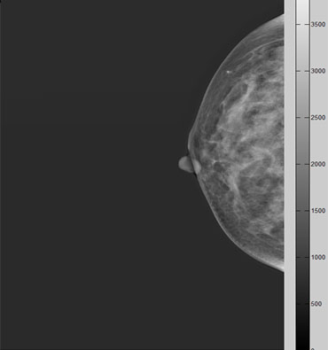
Digital Mammography Image
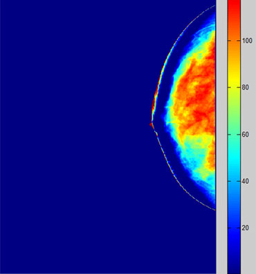
Density Measurement Image
Accuracy of rotation control of C-arm(± 0.5°)
In order to obtain an image for estimating the location of the lesion, the X-ray generator must be accurately moved left and right at a certain angle to obtain the image. The position error of the X-ray generator affects the image for position estimation and cause an error in the estimated position, so the rotational precision of the C-arm directly affects the position estimation. Through mechanism research and clutch improvement to improve the precision of C-arm rotation, the error range has been narrowed down to within ±0.5°

3D Breast Reconstruction Technology
Since conventional mammography takes two-dimensional images composed of flat surface by shooting under the pressure of three-dimensional breast, it is difficult to diagnose of lesions because the suspected lesion is covered with other parts of the breast. To improve this, a 3D image reconstruction technique combining computed tomosynthesis is used to acquire 3D images and diagnose them using slice images.
The advantage of this technique is that a projection image can be obtained with a rotation of 15 to 30 degrees to obtain a stereoscopic image,
thereby greatly reducing the time and X-ray dose and minimizing the pressure on the breast. The pain and fear by breast pressure can be eliminated.
Secured original filtered back projection technology
We constructed a 3D image of projection image using original Filtered Back
projection technique and constructed sliced image for display.
For this purpose, a combination of various filters only for Medi-future was constructed and the algorithm was optimized.

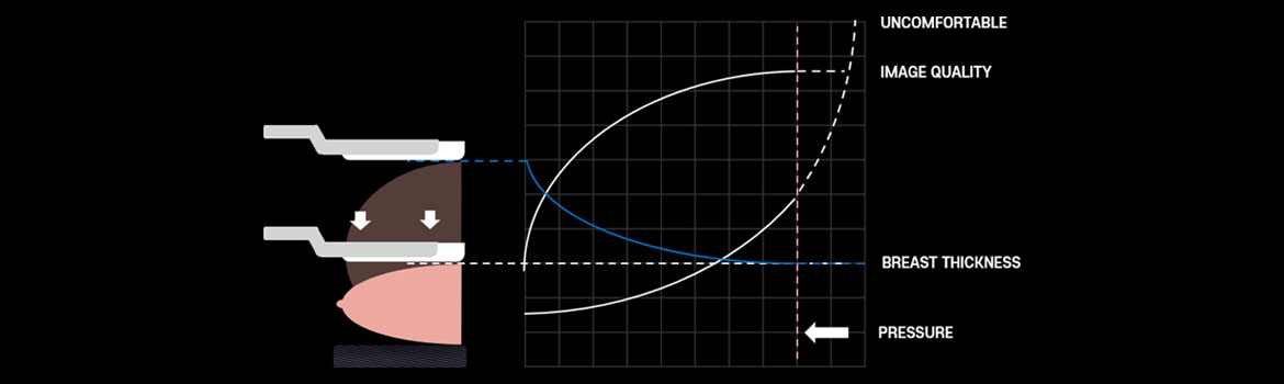
Reduction of pain through a sophisticated motion control
To take a patient's image from a mammography device, the patient's breast is compressed to a maximum of 20kg. In general, the time taken for taking a picture is about 30 seconds and taken twice for each side. The purpose is to fix the breast so that it does not move during the time that it is exposed to the X-ray, and by extracting information on the two-dimensional plane of the three-dimensional breast, it is thinned as much as possible so that much information is expressed on the two- dimensional plane. During the time that the breasts are pressed, the pain caused by the pressure is more individualized, but if you have a tight breasts, you feel more pain. The emotional research on women's breast pressure reduces the pain and measures the distribution of pressure in the breast by using a film, therefore SOUL software can measure the pressure distribution and improves pressure uniformity.
-
1505, Startower, 37, Sagimakgol-ro 62 beon-gil, Jungwon-gu, Seongnam-si, Gyeonggi-do, South Korea 13211
TEL : +82-31-8018-5140
FAX : +82-31-8018-5141
E-mail : sales@medi-future.com
