Technology
Image Processing
Breast Imaging Solutions
MEDI-FUTURE ‘s new image processing algorithms Mammo IP allows you to see the smallest lesions from the breast tissue without losing sight of your patient’s overall needs. You can achieve high end image quality while maintaining low dose.
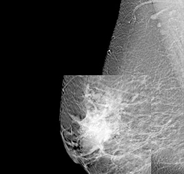 Calcifications
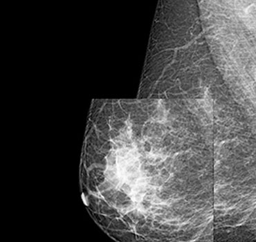 Mass
Mass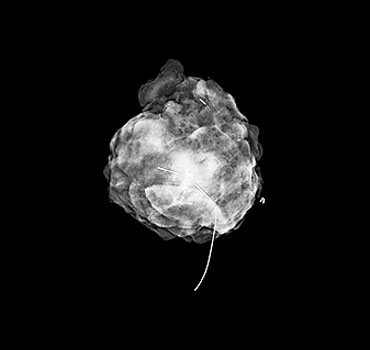 Specimen
Multi-resolution processing
It is a technology that separates images for each frequency in order to process raw image data. It can selectively emphasize or attenuate only the frequency area where there is a lot of important information, so that the image quality can be optimally created. Micro-calcification, a major lesion of the breast cancer, comes from a high frequency area and mass comes from low to mid frequency area, it supports to automatically select and highlight the breast lesion area.
Adaptive dynamic range reduction
It is a technology that converts low dynamic range into high dynamic range, and it is a method of global equalization by reducing the range of change in the breast skin area. Through this algorithm, it is possible to process separately parenchymal tissue, fat and lesion by identifying the intensity due to the difference in X-ray attenuation in the parenchyma and fat in the breast tissue during the X-ray imaging process
Intelligent noise suppression
When reading a breast image, the contrast enhancement process is required to clearly read the parenchyma, skin, muscle and lesions of the breast tissue. This is an algorithm that minimizes the noise generated when the contrast is automatically increased in consideration of the image characteristics of each tissue.
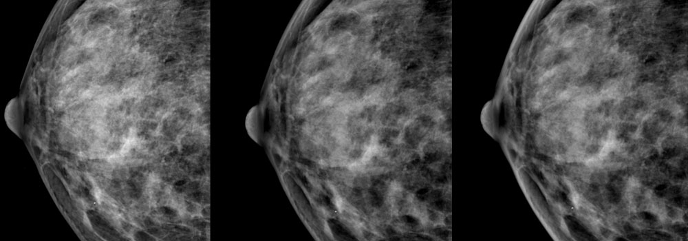
Multi scale local contrast enhancement
A technology that detects the location of intensity distribution of tissue and lesion inside the breast in a breast image. By identifying the location of tissues and lesions in the image, contrast and smoothing are separated by region to visually improve image quality
-
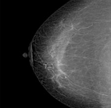
Before Application
-
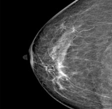
After Application
-
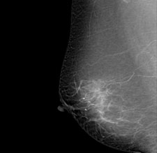
Before Application
-
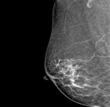
After Application
-
1505, Startower, 37, Sagimakgol-ro 62 beon-gil, Jungwon-gu, Seongnam-si, Gyeonggi-do, South Korea 13211
TEL : +82-31-8018-5140
FAX : +82-31-8018-5141
E-mail : sales@medi-future.com


